The Role of Free-Electron Lasers in Structural Biology and Drug Discovery
Free-electron lasers (FELs) have expanded what researchers can observe in structural biology. By producing extremely bright, ultrashort X-ray pulses, these facilities allow scientists to determine molecular structures and follow biological reactions on timescales ranging from milliseconds to femtoseconds. That combination is especially valuable in drug discovery, where understanding both the shape of a target and its changing interactions with a compound can guide the design of more selective therapies.
X-ray free-electron lasers (XFELs) do not replace synchrotrons, cryo-electron microscopy, nuclear magnetic resonance (NMR), or computational modelling. Instead, they provide a complementary view of biological systems, particularly when samples are radiation-sensitive, available only as microcrystals, or trapped in short-lived functional states.
What are free-electron lasers?
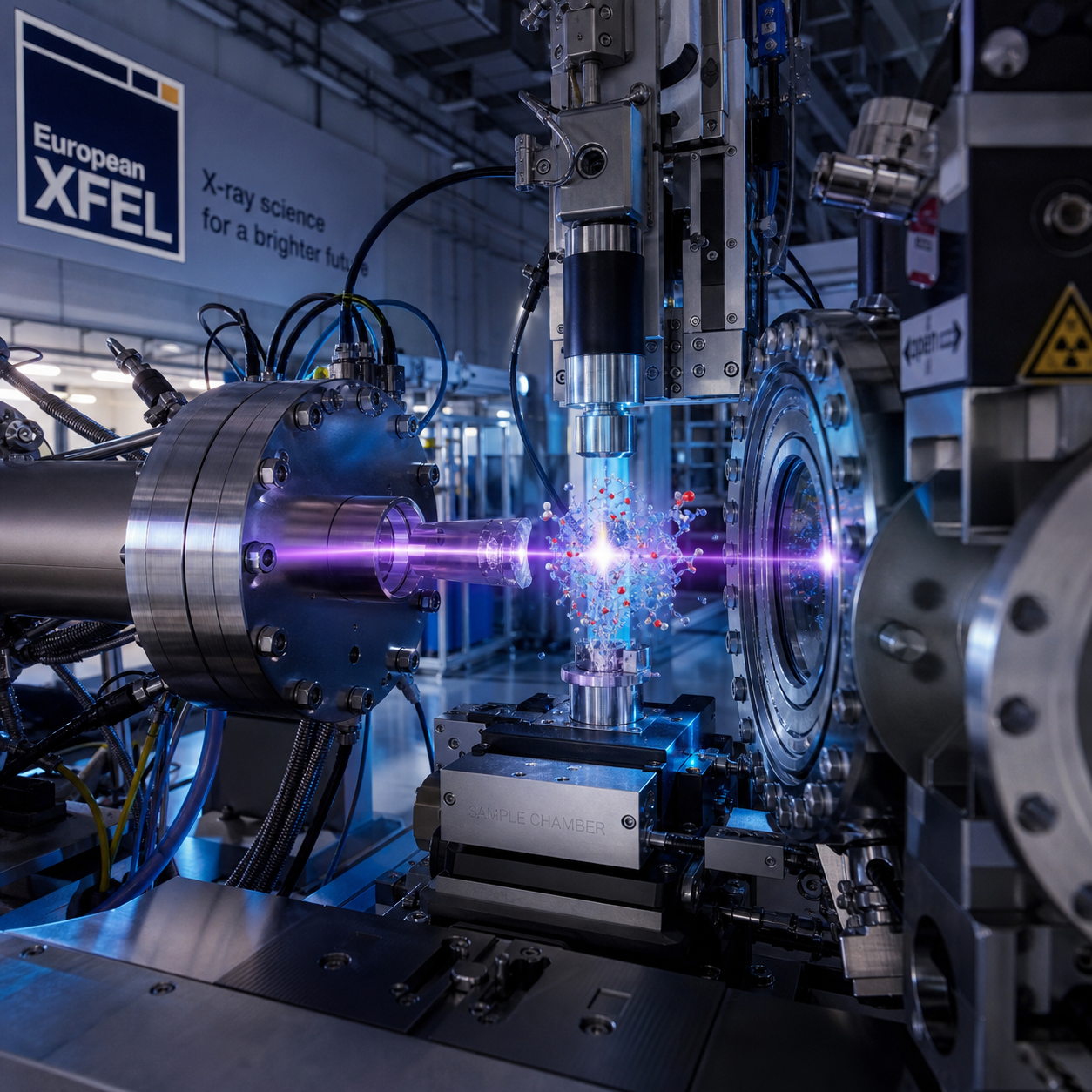
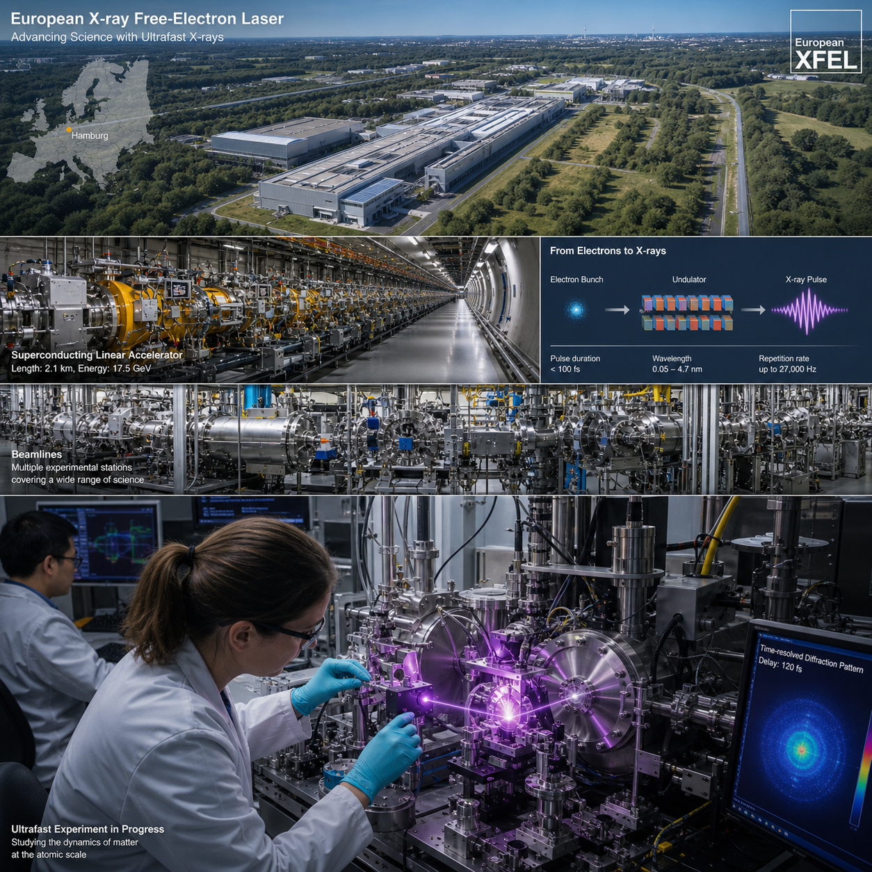
Free-electron lasers generate intense, ultrashort X-ray pulses by accelerating electrons through a long magnetic structure called an undulator. The resulting light has the brightness and timing precision needed to record structural information from biological samples before radiation damage destroys them.
In an XFEL, an electron beam travels through an accelerator and then passes through alternating magnetic fields. These fields make the electrons emit coherent X-rays, producing pulses that can be billions of times brighter than conventional laboratory X-ray sources. Pulse durations may reach the femtosecond range, allowing researchers to capture molecular events almost as they happen.
Two properties matter most for biological research:
- High peak brightness: intense pulses produce measurable diffraction signals from very small crystals or weakly scattering samples.
- Ultrashort duration: a pulse can collect data before substantial radiation damage develops, an approach often described as diffraction before destruction.
- Precise timing: optical, chemical, electrical, or mechanical triggers can synchronize an experiment with a biological reaction.
These capabilities turn an XFEL experiment into more than a search for a static structure. They make it possible to ask when a protein changes shape, how a catalytic intermediate forms, or which molecular contacts appear as a ligand binds.
How FELs advance structural biology
FELs advance structural biology by revealing high-resolution macromolecular structures and transient conformations that are difficult to capture with conventional methods. Researchers can investigate proteins, enzymes, membrane proteins, viruses, and large molecular assemblies under conditions closer to their functional state.
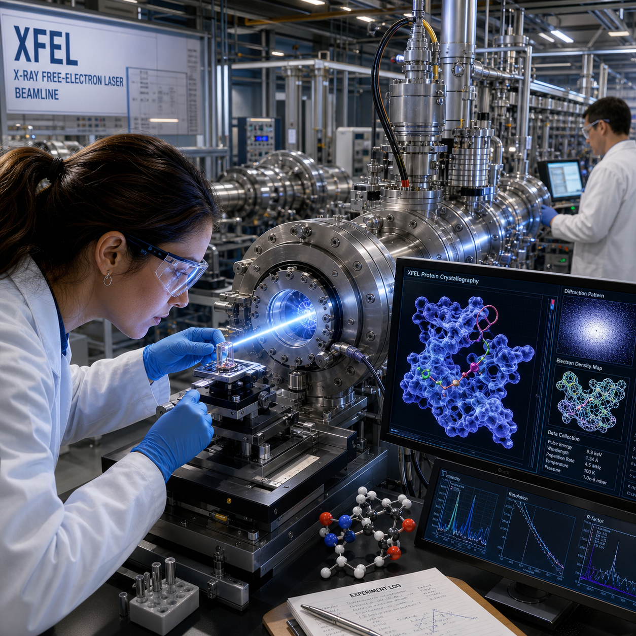
Protein crystallography remains central to many XFEL studies. A crystal scatters X-rays into a diffraction pattern, and computational methods convert those measurements into an electron-density map. From that map, scientists build an atomic model of the protein or molecular complex.
Traditional crystallography often requires a relatively large, well-ordered crystal and exposure times long enough for radiation damage to accumulate. XFEL pulses change the balance. A stream of small crystals can be exposed one at a time, with each pulse recording a diffraction image before the individual crystal is seriously damaged.
This approach helps address several persistent structural biology problems:
- Small or fragile crystals: samples that are unsuitable for standard rotation experiments may still produce useful snapshots.
- Radiation-sensitive chemistry: metal centres, redox enzymes, and catalytic cofactors can be examined with reduced damage during each exposure.
- Conformational heterogeneity: ensembles of structures can reveal alternative states rather than forcing a sample into one dominant model.
- Membrane and complex proteins: difficult targets can be studied when microcrystallisation or specialised sample delivery succeeds.
XFEL data still require careful interpretation. Resolution, completeness, crystal quality, indexing accuracy, and model validation remain important. A powerful beam does not compensate for poorly prepared samples or an experiment that lacks a clear biological question.
Key techniques enabled by XFEL facilities
XFEL facilities enable serial femtosecond crystallography, time-resolved crystallography, microcrystallography, and other approaches for challenging biological samples. These techniques convert many short exposures into structural information about static and moving molecular systems.
Serial femtosecond crystallography
Serial femtosecond crystallography (SFX) streams thousands of crystals through an XFEL beam. Each crystal contributes one or more still diffraction patterns, which are combined computationally to reconstruct a three-dimensional structure.
SFX avoids the need to rotate a single crystal through many angles. It can therefore work with crystals that are too small, radiation-sensitive, or numerous to mount individually. Sample delivery may use liquid jets, fixed targets, viscous media, or other systems selected according to crystal size and concentration.
Time-resolved crystallography
Time-resolved crystallography introduces a trigger before the X-ray pulse records the structural response. Light activation, rapid mixing, temperature changes, ligand delivery, or enzymatic turnover can initiate the reaction. By varying the delay between trigger and probe, researchers build a molecular movie from a series of structural snapshots.
The time resolution depends on the trigger, sample environment, pulse timing, and reaction synchronisation. A femtosecond pulse does not automatically mean that every experiment resolves femtosecond biology; the sample must respond uniformly and the reaction must be initiated precisely.
Microcrystallography and difficult samples
Microcrystallography focuses the beam on crystals that may be only a few micrometres across. XFEL beamlines also support specialised sample environments for membrane proteins, enzymes, photoreceptors, and radiation-sensitive complexes. In some cases, complementary data from synchrotron crystallography, cryo-EM, spectroscopy, or molecular dynamics provide the context needed to interpret the FEL structure.
Applications in drug discovery
FELs support drug discovery by showing how candidate molecules occupy binding sites, alter protein structure, and interact with targets over time. This information strengthens structure-based drug design, fragment screening, target validation, and optimisation of protein–ligand interactions.
In a conventional structure-based workflow, researchers use an atomic protein model to identify a pocket and design compounds that fit it. XFEL data can add details that a single static structure may miss, including alternate side-chain positions, water networks, induced-fit movements, and short-lived binding modes.
Fragment screening
Fragment screening tests small chemical building blocks against a target. Because fragments are compact, they often bind weakly but reveal efficient interactions that medicinal chemists can elaborate into larger molecules. Serial crystallography can examine many soaked or co-crystallised crystals, making it useful for mapping fragment-binding sites across a protein surface.
The practical value lies in the quality of the structural evidence. A fragment hit supported by clear electron density, sensible chemistry, and reproducible occupancy gives a stronger starting point than a screening signal without a defined binding mode. XFEL measurements may also distinguish between multiple fragments occupying nearby subsites.
Protein–ligand interactions and target validation
XFEL structures help researchers assess hydrogen bonds, hydrophobic contacts, metal coordination, solvent-mediated interactions, and pocket rearrangements. These observations can explain why a compound is selective, why resistance mutations weaken binding, or why a biochemical inhibitor fails to engage the target in its functional conformation.
For target validation, structural results become most informative when combined with biochemical assays, cellular data, medicinal chemistry, and computation. A high-resolution complex confirms a physical interaction, but it does not by itself establish efficacy, safety, pharmacokinetics, or clinical potential.
From molecular snapshots to dynamic mechanisms
Time-resolved FEL experiments reveal conformational changes, reaction pathways, and transient intermediates that connect molecular structure with biological function. For drug research, this dynamic information can expose regulatory states and binding opportunities that remain hidden in equilibrium structures.
Many proteins behave like flexible machines rather than rigid locks. Enzymes open and close access channels, receptors shift between signalling states, and transporters alternate between inward- and outward-facing conformations. A ligand may stabilise one state, block a transition, or redirect the reaction pathway.
Researchers can use a pump–probe design to study these events. A pump initiates the biological process, while a delayed X-ray pulse probes the structure. Repeating the measurement at different delays produces a sequence of time points. The resulting models can identify intermediate conformations, moving cofactors, protonation-linked changes, or rearrangements around a catalytic site.
This matters for therapeutic development because a transient state may offer a selective binding pocket. It may also explain mechanism-based inhibition, allosteric regulation, or the molecular basis of drug resistance. Still, dynamic interpretation requires caution. Data may represent a mixture of states, and the reaction conditions in a crystal may differ from those in a living cell.
A useful working model is the structure–motion–chemistry chain: first define the atomic structure, then identify the motion or state transition, and finally connect that transition to chemical activity or ligand binding. FEL experiments are strongest when they answer all three questions with complementary measurements.
The role of European FEL infrastructure and collaboration
The European FEL network gives researchers access to specialised beamlines, sample environments, instrumentation, and multidisciplinary expertise that individual laboratories usually cannot maintain alone. Shared infrastructure also enables cross-border projects linking structural biology, chemistry, physics, computation, and pharmaceutical research.
European facilities such as the European XFEL and other regional free-electron laser centres provide capabilities that must be coordinated carefully. Beamtime proposals typically need a defined scientific question, a credible sample plan, appropriate controls, and a realistic data-analysis strategy. Access arrangements vary by facility and programme, so researchers should consult the relevant user office and current call documentation rather than assume a single route.
The infrastructure contributes at several levels:
- Beamline science: optics, detectors, timing systems, and experimental control support high-quality measurements.
- Sample environments: injectors, fixed targets, mixing devices, light-activation systems, and cryogenic or ambient-condition setups connect the beam to biological questions.
- Expert teams: crystallographers, beamline scientists, biochemists, data scientists, and computational researchers help translate a proposal into a workable experiment.
- Data analysis: indexing, integration, merging, phasing, refinement, and time-series analysis require robust pipelines and substantial computing resources.
Collaboration is especially important for drug discovery programmes, where a structure may need to be linked quickly to compound synthesis and biological testing. European research networks can reduce duplication, share protocols, and make specialised knowledge available to groups that are new to XFEL methods.
Authoritative technical context on free-electron lasers and their applications is also available through the European synchrotron and research infrastructure community, which illustrates how photon facilities operate as complementary parts of modern structural biology rather than isolated technologies.
Challenges and future opportunities
FEL experiments remain technically demanding because they require suitable samples, precise delivery, high-throughput data processing, and scarce facility access. Future progress will depend on automation, better sample economy, integrated analysis, and closer links between FEL measurements and drug-development workflows.
Sample preparation is often the first bottleneck. Producing thousands of uniform microcrystals can consume significant protein, while viscous injectors or fixed targets may impose different concentration and viscosity requirements. Researchers should test crystallisation, diffraction, delivery, and reaction triggering well before a scheduled experiment.
Data volume creates a second challenge. A single run can generate large numbers of detector images, many of which contain weak or incomplete diffraction. Automated pipelines speed processing, but scientists still need to inspect hit rates, resolution limits, indexing ambiguity, radiation effects, and model bias.
Common planning mistakes include:
- Choosing XFELs solely because they are powerful: the method may add little value if a stable, well-diffracting crystal already answers the biological question at a synchrotron.
- Underestimating sample consumption: serial experiments can require more material than a single-crystal measurement, depending on delivery efficiency and hit rate.
- Assuming a time-resolved design guarantees a molecular movie: poor triggering or asynchronous reactions can blur intermediate states.
- Leaving analysis until after beamtime: a defined processing plan and rapid feedback are essential for deciding whether additional data are needed.
Emerging opportunities include robotic crystal screening, adaptive data collection, improved timing diagnostics, machine-learning-assisted analysis, and integration with cryo-EM maps, NMR restraints, spectroscopy, molecular dynamics, and medicinal chemistry. These developments could make FEL studies more routine, although cost, access, and experimental complexity will continue to shape when the method is justified.
Frequently asked questions
What is an X-ray free-electron laser used for in structural biology?
An X-ray free-electron laser is used to determine high-resolution structures and follow changes in proteins, enzymes, membrane systems, and molecular complexes. Its ultrashort pulses are particularly useful for radiation-sensitive samples and time-resolved experiments.
How do FELs differ from conventional synchrotron sources?
FELs deliver much shorter pulses with very high peak brightness, while synchrotrons provide highly reliable, tunable, and often more accessible X-ray beams for routine crystallography and spectroscopy. The two source types are complementary.
Can XFELs study protein–drug interactions?
Yes. XFELs can resolve protein–ligand binding modes, support fragment screening, and investigate conformational changes caused by inhibitors or activators. Results should be combined with biochemical, cellular, and computational evidence.
What is serial femtosecond crystallography?
Serial femtosecond crystallography is a method in which many microcrystals are exposed sequentially to XFEL pulses. Each crystal contributes diffraction data before significant radiation damage, and the images are merged into a structural model.
How can researchers access European FEL facilities?
Researchers generally apply through facility user offices, proposal calls, national or international access programmes, and collaborative projects. The exact process, eligibility rules, and review criteria differ, so teams should check the current information published by the relevant European FEL facility.
Free-electron lasers broaden structural biology by connecting atomic detail with molecular motion. In drug discovery, that connection can reveal binding modes, transient pockets, catalytic intermediates, and mechanisms of selectivity or resistance. Their greatest value emerges when XFELs are used alongside synchrotrons, cryo-EM, NMR, spectroscopy, and computation as part of a coordinated European research infrastructure. The result is a more complete picture of how biological targets work and how therapeutic molecules can influence them.