Exploring the Unique Capabilities of European XFEL Facilities

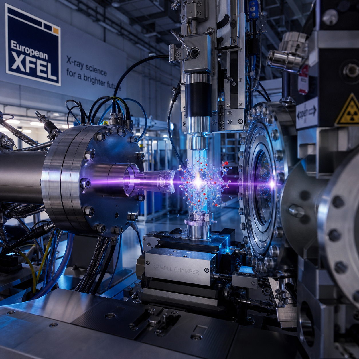
European X-ray free-electron laser facilities give researchers access to flashes of X-ray light that are exceptionally bright, coherent, and brief. These pulses allow scientists to examine atomic structures, chemical reactions, magnetic states, and extreme forms of matter while they change.
The European XFEL network connects large-scale free-electron laser laboratories with specialized instruments, advanced detectors, and international research teams. Its value lies in the combination of pulse properties and experimental infrastructure: the source creates the X-rays, while beamlines and experimental stations turn them into measurements that answer precise scientific questions.
What Makes X-ray Free-Electron Lasers Different?
X-ray free-electron lasers produce intense, coherent X-ray pulses that can last for femtoseconds. This combination of brightness, coherence, ultrashort duration, and high repetition rate enables experiments that conventional light sources often cannot perform in the same way.
An X-ray free-electron laser, or XFEL, accelerates a beam of electrons to very high energy and passes it through an array of magnets called an undulator. The electrons emit X-ray radiation that becomes organized into powerful laser-like pulses. Unlike a conventional optical laser, the process does not rely on a traditional gain medium such as a crystal or gas.
Three properties matter especially:
- Ultrashort pulse duration: Femtosecond pulses can freeze a rapid stage of molecular motion, much as a very fast camera captures a moving object without ordinary motion blur.
- High brightness: A large number of X-ray photons arrive in a short interval, providing enough signal to study tiny samples or weak scattering processes.
- Coherence: Coherent X-ray pulses have coordinated wave properties that support phase-sensitive imaging and detailed studies of ordered structures.
These advantages come with practical demands. A pulse can damage or destroy a sample, so experiments often use fresh material for every exposure. The scientific design therefore includes sample delivery, timing synchronization, radiation shielding, data acquisition, and rapid analysis from the beginning.
Researchers can consult the European XFEL facility website for current information about instruments, scientific areas, and user access.
European XFEL’s Facility Architecture and Research Infrastructure
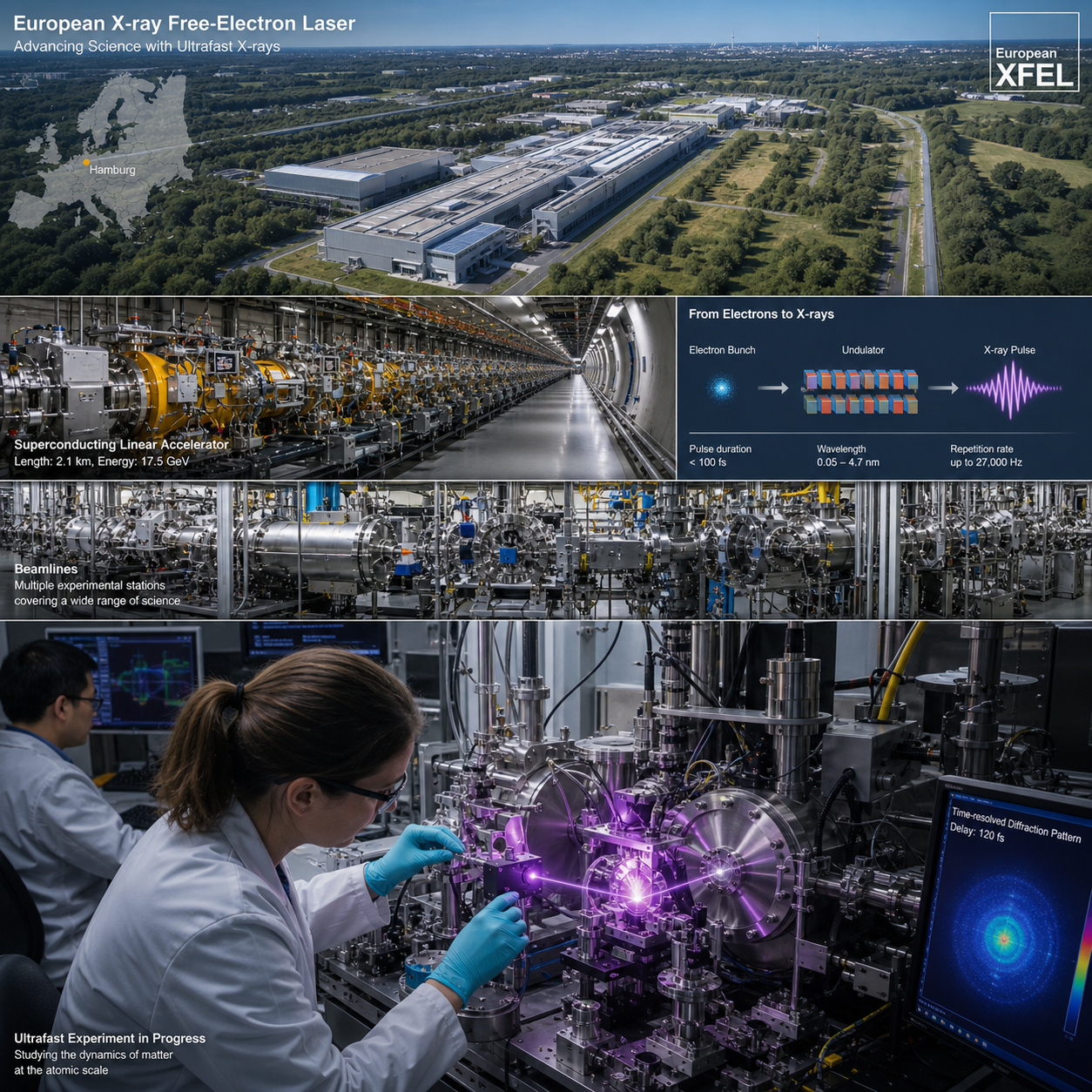
European XFEL combines a superconducting accelerator, photon systems, beamlines, experimental stations, and large-scale detectors into one research infrastructure. This architecture allows scientists to move from electron acceleration to usable X-ray measurements within a coordinated facility.
The accelerator generates the electron bunches that produce the X-ray pulses. Photon systems then transport, focus, split, or condition the radiation for different experiments. Long beamlines guide the X-rays to experimental stations, where researchers introduce samples and record scattering, diffraction, spectroscopy, or imaging signals.
From source to measurement
- Accelerator: Produces high-energy electron bunches with carefully controlled timing and properties.
- Undulators and photon transport: Convert electron motion into X-ray pulses and deliver them to instruments.
- Beamlines: Shape and direct the beam while preserving the conditions needed for a particular experiment.
- Experimental stations: Combine sample environments, diagnostics, timing tools, and detectors.
- Detectors and computing: Capture rapid streams of data and support reconstruction, correction, and interpretation.
This integrated design is important because an XFEL experiment rarely consists of simply placing a sample in an X-ray beam. Researchers may need a liquid jet, a vacuum chamber, a high-pressure cell, a laser system, a cryogenic environment, or a synchronized particle source. Facility scientists and instrument teams help match those requirements to available capabilities.
Capturing Matter in Motion
XFEL experiments capture matter in motion by combining femtosecond time resolution with controlled excitation and X-ray probing. The approach reveals how atomic arrangements change after a reaction, phase transition, light pulse, or other trigger.
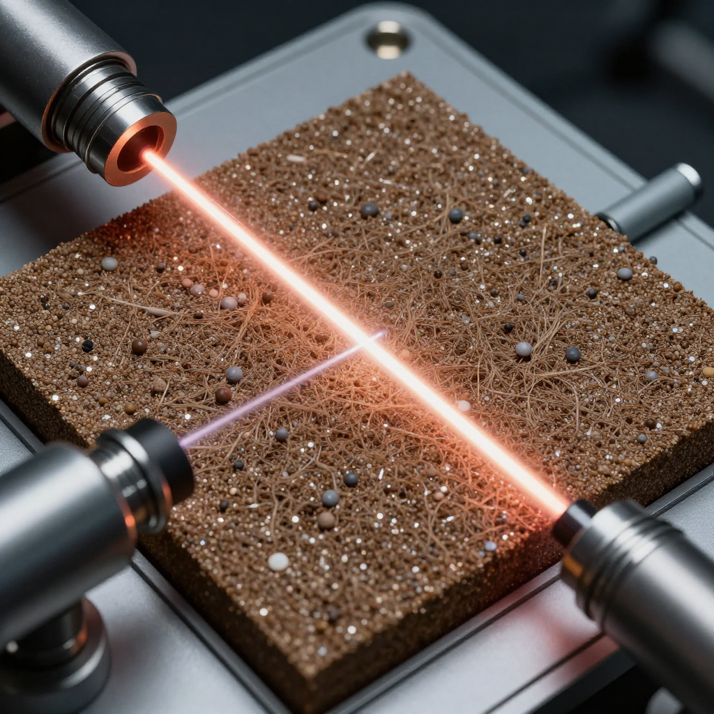
In a common pump–probe experiment, a pump pulse first stimulates the sample. It might be an optical laser, an electrical signal, a chemical reaction, or a shock wave. After a controlled delay, an XFEL pulse probes the sample. Repeating the measurement at many delays creates a sequence of structural snapshots.
For example, researchers can ask whether a molecule changes shape before a bond breaks, how a semiconductor reorganizes after absorbing light, or when magnetic order begins to shift. The X-ray pulse does not simply provide a final image; it supplies a time-stamped measurement of structural or electronic change.
Time resolution depends on more than the nominal pulse length. Synchronization between pump and probe, arrival-time diagnostics, sample thickness, detector response, and the physical process itself all affect the effective resolution. A shorter pulse is useful only when the rest of the experiment can preserve that timing advantage.
Key Research Applications Across Disciplines
XFEL facilities support research in structural biology, chemistry, materials science, condensed matter, plasma physics, and related fields. Across these disciplines, the common goal is to connect structure and dynamics at very small scales.
Structural biology
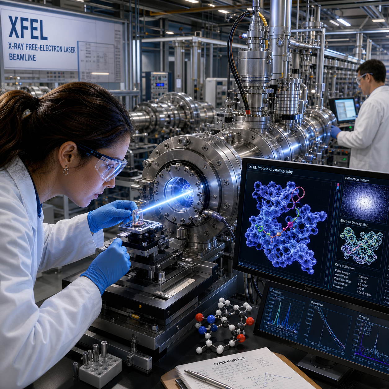
In structural biology, scientists use diffraction and imaging methods to investigate proteins, enzymes, viruses, and other biological assemblies. XFEL pulses are particularly valuable when radiation damage limits conventional measurements or when a sample exists only in small crystals. Structural information can be collected rapidly, sometimes before the sample is significantly altered by the radiation.
Chemistry and catalysis
Chemists use time-resolved X-ray diffraction and spectroscopy to follow intermediates in chemical reactions. These measurements can reveal changes in oxidation state, coordination geometry, or molecular structure that may be invisible in a measurement taken only before and after the reaction.
Materials science and condensed matter
Materials researchers study phase transitions, strain, defects, superconducting behavior, charge order, and magnetic dynamics. Pump–probe experiments can link an external stimulus to changes in lattice structure or electronic organization over femtoseconds to longer timescales.
Plasma and extreme conditions
High-intensity lasers and XFEL pulses can create or diagnose matter at extreme temperature, pressure, or density. These studies contribute to plasma physics and laboratory astrophysics, although the required equipment and safety arrangements vary substantially between experimental stations.
XFELs do not replace synchrotrons, electron microscopes, neutron sources, or laboratory lasers. Each tool measures different signals under different conditions. The strongest projects often combine facilities rather than treating one source as universally superior.
Advanced Experimental Techniques and Instruments
Advanced XFEL techniques translate coherent X-ray pulses into structural, chemical, and imaging information. The appropriate method depends on the sample, the timescale of interest, the expected signal, and whether the sample can survive repeated exposure.
- Serial femtosecond crystallography: A stream of microcrystals passes through the beam, and each crystal contributes a diffraction pattern before radiation damage becomes dominant. Computational methods combine thousands or millions of patterns into a structure.
- Time-resolved diffraction: Measures how crystal lattices or ordered materials change after a pump event.
- Spectroscopy: Probes electronic states, chemical environments, and elemental behavior through energy-dependent X-ray interactions.
- Coherent imaging: Uses the phase relationships in coherent X-ray pulses to reconstruct features that may not be visible through conventional absorption contrast.
- Single-particle studies: Seek structural information from isolated particles or non-crystalline ensembles, supported by careful sample delivery and substantial computational analysis.
Technique selection should follow the measurement question. If the goal is a molecular structure, diffraction may be central. If the question concerns electronic rearrangement, spectroscopy could provide the more direct signal. A sophisticated instrument cannot compensate for an unclear observable or insufficient sample statistics.
The Role of the European XFEL Network
The European XFEL network expands research capacity by connecting complementary facilities, instruments, expertise, and international collaborations. European facilities may differ in pulse characteristics, repetition rate, photon-energy range, sample environments, detector systems, and specialist instruments.
This complementarity gives research teams more than one route to a scientific objective. One facility may offer a particularly suitable timing configuration, while another provides a specialized detector, high-pressure environment, or established expertise in serial crystallography. Shared knowledge also helps researchers transfer sample-preparation methods, analysis workflows, and experimental designs between laboratories.
International access is a defining feature of these free-electron laser laboratories. Experiments commonly involve principal investigators, facility scientists, sample-delivery specialists, laser experts, data scientists, and instrument operators. The collaboration is practical: successful beamtime depends on integrating many technical decisions under strict scheduling and safety constraints.
Researchers should compare facilities by experimental fit rather than reputation alone. Useful questions include:
- Does the source provide the required photon energy and pulse structure?
- Can the instrument handle the sample form, environment, and delivery method?
- Are the detector, timing tools, and data systems appropriate for the expected signal?
- Does the facility team have experience with the proposed measurement?
How Researchers Engage with XFEL Facilities
Researchers usually engage with XFEL facilities by selecting a suitable instrument, submitting a scientifically justified proposal, developing an experimental plan, and carrying out the work with facility scientists. Exact access rules and proposal cycles differ between facilities, so applicants should check the current user-office guidance.
- Define the measurement: State what quantity must be observed, such as a transient structure, diffraction pattern, chemical state, or image.
- Choose the facility and instrument: Match photon energy, pulse timing, repetition rate, sample environment, detector, and technique to the question.
- Prepare a realistic proposal: Explain the scientific case, sample numbers, controls, expected signal, analysis plan, and reason XFEL capability is necessary.
- Plan feasibility: Address sample delivery, radiation safety, laser synchronization, cryogenics, vacuum, travel, and contingency measurements.
- Conduct and analyze the experiment: Work with beamline scientists during setup and collect enough metadata to make later data interpretation reliable.
A common mistake is proposing an ambitious time-resolved experiment without demonstrating that the sample can be prepared, delivered, and measured repeatedly. Another is underestimating analysis: high-repetition-rate experiments can generate large datasets, so storage, calibration, reconstruction, and computing should appear in the plan.
Strong proposals make a clear chain of reasoning: scientific question, measurable signal, facility capability, feasible method, expected contribution. That chain helps reviewers distinguish an experiment that genuinely requires an XFEL from one that another laboratory technique could answer more efficiently.
Frequently Asked Questions About European XFEL Facilities
What is an X-ray free-electron laser?
An X-ray free-electron laser is a large-scale source that uses accelerated electrons and undulators to generate intense, coherent X-ray pulses. The pulses can be short enough to resolve ultrafast structural and electronic dynamics.
What makes European XFEL facilities unique?
They combine advanced X-ray pulse properties with specialized beamlines, experimental stations, detectors, sample environments, timing systems, and international scientific support. The full infrastructure enables measurements that depend on both the source and the instrument.
Which research fields use XFEL experiments?
Major fields include structural biology, chemistry, catalysis, materials science, condensed matter physics, plasma physics, nanoscience, and ultrafast science. Projects vary from protein structure determination to studies of phase transitions and matter under extreme conditions.
How can researchers apply for XFEL beamtime?
Researchers generally submit a proposal through the relevant facility’s user-access system. The proposal should identify the scientific question, explain why XFEL pulses are needed, describe the sample and method, and demonstrate experimental feasibility.
What can ultrafast XFEL pulses reveal?
Ultrafast XFEL pulses can reveal how atomic positions, molecular structures, electronic states, magnetic order, and material phases evolve after a controlled stimulus. They provide snapshots that help connect rapid processes to their underlying mechanisms.
European XFEL facilities are best understood as coordinated scientific ecosystems rather than interchangeable X-ray sources. Their distinctive capabilities emerge when coherent X-ray pulses, femtosecond timing, specialized instruments, robust detectors, and expert collaboration are designed around one well-defined question: how does matter change, and what happens first?