Advances in Ultrafast Science Enabled by Free-Electron Lasers

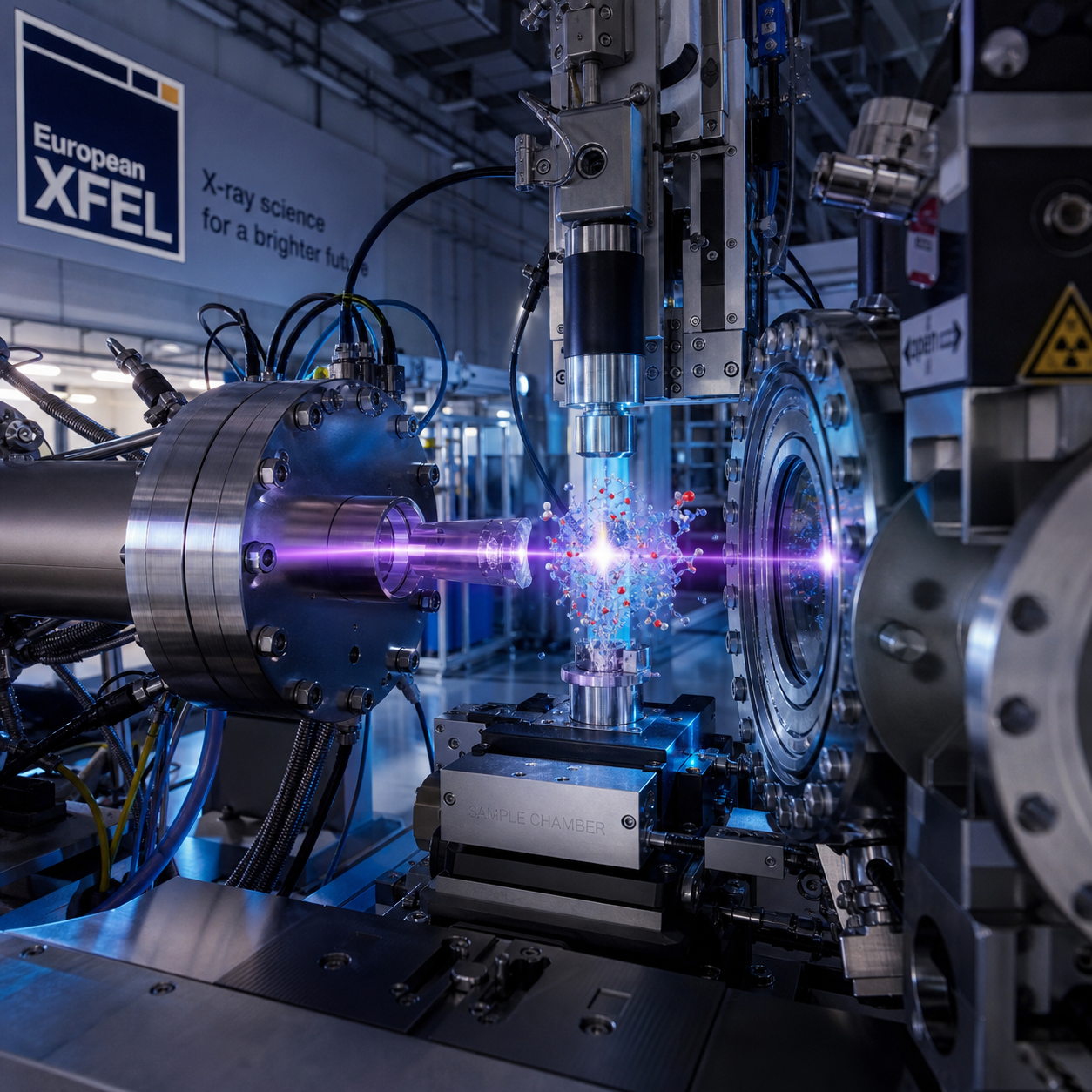
Advances in ultrafast science enabled by free-electron lasers are giving researchers new ways to study matter while electronic, atomic and molecular changes unfold. These facilities combine femtosecond or shorter light pulses with tunable photon energies, high brightness and intense X-ray beams, allowing experiments that were previously limited by time resolution, radiation sensitivity or insufficient structural information.
The result is a more detailed view of chemical reactions, materials, biological structures, plasma and quantum systems. FEL experiments do not produce a universal camera-like recording of matter in real time; instead, carefully designed measurements reconstruct dynamics from signals collected at controlled time delays.
Why ultrafast science needs advanced light sources
Ultrafast science examines processes that occur from femtoseconds to attoseconds, often across atomic and molecular length scales. Conventional measurement methods can average over these changes, making it difficult to determine which event happened first or how energy moved through a system.
A femtosecond is 10-15 seconds. Electron rearrangement, bond formation and energy transfer can begin within this interval, while nuclear motion often follows on a slightly longer timescale. Attosecond pulses target even faster electron dynamics. At the same time, researchers may need ångström-scale structural information, especially when atoms shift by only a fraction of a nanometre.
This creates a demanding measurement problem. The probe must be short enough to freeze the relevant motion, energetic enough to access the required electronic or structural signal, and bright enough to produce useful data from a small sample. It must also arrive with reliable timing relative to the event being studied.
Visible and infrared lasers remain essential for initiating reactions and driving materials. However, they generally do not provide the same direct access to elemental composition, local electronic states or atomic structure as short-wavelength X-rays. The combination of an optical pump and an X-ray probe therefore expands what time-resolved experiments can determine.
How free-electron lasers enable new measurements
A free-electron laser generates intense, tunable light when a relativistic electron beam passes through a periodic magnetic structure called an undulator. In an X-ray free-electron laser, or XFEL, the electron beam interacts collectively with the emitted radiation, producing highly bright pulses at wavelengths suited to atomic-scale research.
Unlike a conventional laser medium, an FEL uses accelerated electrons as its source. The electron bunch travels through the undulator, where its motion creates radiation that can become coherent and extremely intense. Large accelerator-based facilities then deliver the pulses to specialised experimental stations.
- Ultrashort duration: Femtosecond pulses can separate stages of a reaction, while emerging attosecond methods address electron motion.
- Tunable photon energy: Researchers can select wavelengths for spectroscopy, diffraction, imaging or element-sensitive measurements.
- High brightness: A large number of photons in a short pulse supports measurements from dilute, small or transient samples.
- Coherent X-rays: Spatial and temporal coherence enable phase-sensitive imaging, nanoscale studies and advanced scattering approaches.
- Single-pulse capability: Some experiments collect information before radiation damage destroys the individual sample particle.
These capabilities involve trade-offs. High pulse intensity can improve signal but also cause nonlinear effects, sample damage and complex interpretation. Pulse duration at the source does not automatically equal the time resolution of an experiment; timing jitter, detector response, sample thickness and instrument geometry all contribute to the effective resolution.
Technical background on FEL principles and accelerator-based light sources is available through the U.S. Department of Energy Office of Science.
From snapshots to molecular movies
Pump–probe experiments use one pulse to start a process and a delayed pulse to measure its state. Repeating the measurement at different delays produces a time-dependent dataset from which electronic, atomic or molecular dynamics can be reconstructed.
In a typical experiment, an optical or infrared pump excites a sample. After a controlled delay, an X-ray FEL pulse probes the changed state. The detector records diffraction, absorption, emission or photoelectron signals. Researchers then compare the excited-state measurement with a reference and analyse how the signal evolves with delay.
What the probe can reveal
- Time-resolved spectroscopy can follow changes in oxidation state, charge distribution, electronic occupancy and local chemical bonding.
- Time-resolved diffraction can detect lattice motion, phase transitions and structural rearrangements.
- Small-angle scattering and imaging can track nanoscale shape changes, aggregation or domain formation.
- Photoelectron measurements can connect energy-resolved electronic signals with the timing of charge transfer.
A molecular movie is therefore a reconstruction assembled from many measurements, models and constraints. The experiment must control the pump fluence, delay calibration and sample conditions carefully. If the sample changes between shots or the timing drifts, apparent dynamics may reflect experimental instability rather than the underlying science.
Breakthrough applications across research fields
FELs advance research wherever structure, electronic state and dynamics must be measured together. Their applications span chemistry, materials science, structural biology, plasma physics, high-energy-density science and quantum systems.
Chemical reactions and energy conversion
Ultrafast X-ray spectroscopy can follow catalytic intermediates, charge-transfer reactions and photochemical pathways. Researchers can ask when a bond weakens, where an electron localises and how a transient species evolves before returning to a stable state. This information helps test reaction mechanisms that steady-state measurements cannot distinguish.
Materials and condensed matter
In condensed matter, FEL pulses probe phase transitions, magnetic order, superconducting responses and light-induced changes in electronic structure. Time-resolved diffraction can link a change in conductivity or magnetism to a specific lattice distortion. The intense probe also makes it possible to study metastable states that exist only briefly after excitation.
Structural biology
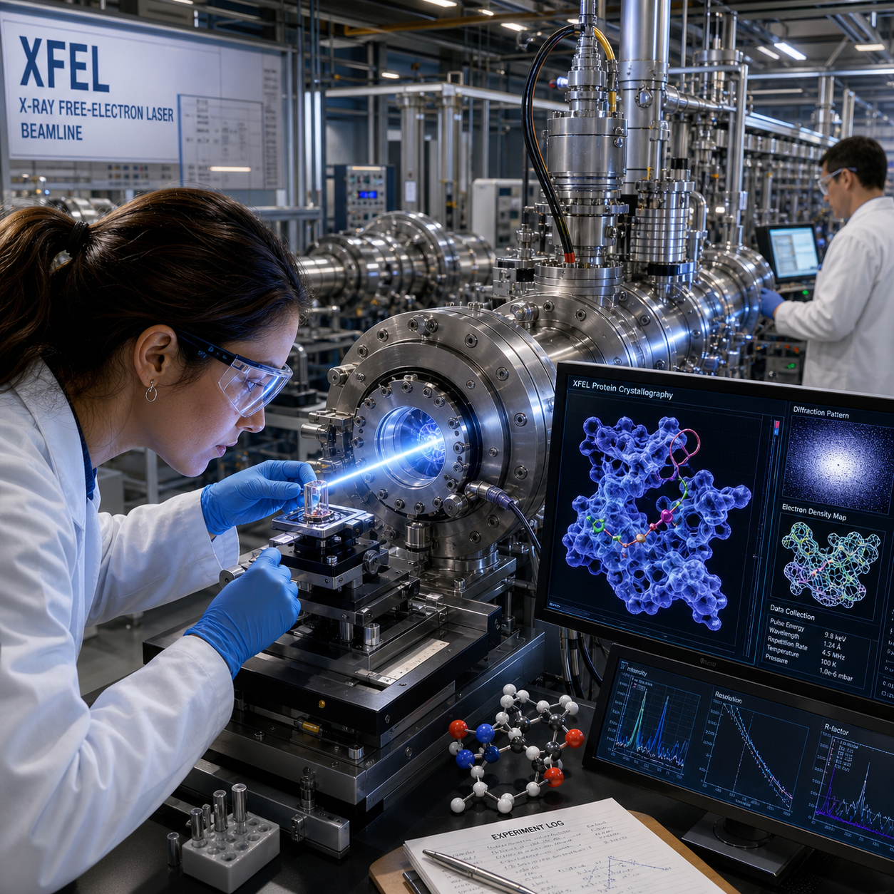
Serial femtosecond crystallography uses streams of microcrystals and collects diffraction before radiation damage fully develops in each crystal. This approach can resolve conformational changes in proteins and enzymes, including reaction pathways initiated by light or substrate binding. Sample consumption, crystal quality and data-processing requirements remain significant constraints.
Plasma and high-energy-density science
Short X-ray pulses can diagnose matter at extreme temperature and pressure, including warm dense matter and laser-driven plasmas. Spectroscopy provides information about ionisation, density and electronic states during conditions that are difficult to reproduce or measure with slower probes.
Quantum and nanoscale systems
FELs also investigate collective excitations, nanoscale magnetism, charge-density waves and nonequilibrium quantum states. Coherent scattering can reveal spatial correlations, while ultrashort timing distinguishes competing relaxation pathways.
The role of European FEL infrastructure and collaboration
European FEL infrastructure supports ultrafast science through multiple large-scale facilities, shared expertise and coordinated access. The European FEL network is not one instrument; it connects distinct facilities, beamlines, detectors, sample environments and scientific communities.
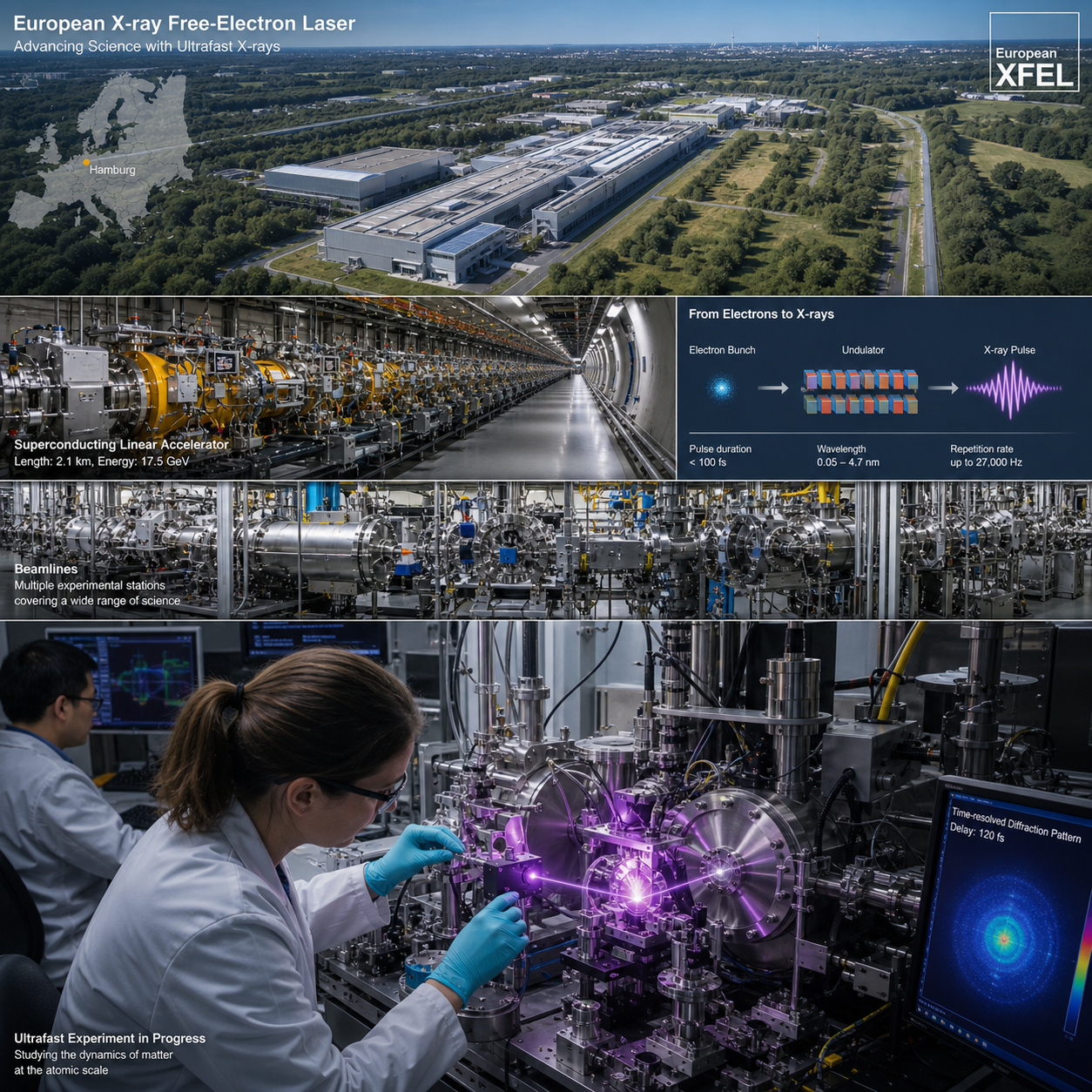
Facilities such as the European XFEL in Germany, FERMI in Italy, SwissFEL in Switzerland and other European accelerator-based light sources provide different photon-energy ranges, pulse properties and experimental capabilities. Some specialise in hard X-rays, others in soft X-rays, seeded coherence or particular spectroscopy and imaging methods.
Access typically depends on a peer-reviewed proposal, scientific feasibility and beamline suitability. Successful teams often combine expertise in accelerator science, instrumentation, chemistry, biology, materials preparation, data analysis and theory. This multidisciplinary model matters because an FEL experiment is an integrated system rather than a simple measurement service.
European collaboration also supports instrument development, training, remote planning and reproducibility practices. Researchers can compare results across facilities, use complementary photon sources and build larger datasets. The compromise is logistical complexity: proposals may require long lead times, specialised sample shipment, safety reviews and substantial preparation before a limited beamtime allocation.
Designing successful FEL experiments
To design a successful FEL experiment, define the dynamic question first, then match the probe, sample and timing system to that question. A strong proposal explains what changes, on what timescale, and which observable can distinguish competing explanations.
- Specify the scientific event. Define whether the target is charge transfer, bond rearrangement, lattice motion, phase switching or another process. Estimate its timescale rather than requesting the shortest pulse automatically.
- Choose photon energy and method. Select absorption edges for element-specific spectroscopy, suitable energies for diffraction, or higher energies when penetration and structural resolution are priorities.
- Plan the excitation and timing. Determine pump wavelength, fluence, spot size and repetition strategy. Include timing-jitter measurement, arrival-time diagnostics and a delay range that covers both early and late dynamics.
- Engineer sample delivery. Decide whether the experiment needs a fixed target, liquid jet, aerosol, tape drive, microcrystal stream or another approach. Confirm concentration, flow rate, refresh rate and environmental control.
- Manage radiation effects. Measure whether the sample changes during exposure. Lowering the dose may protect the sample but reduce signal, so fresh material, scanning strategies or serial delivery may be necessary.
- Build analysis into the plan. Define calibration standards, dark measurements, reference states, metadata and statistical checks before beamtime. Include models that can separate instrument response from physical dynamics.
Common mistakes include choosing a facility because it is familiar rather than because its photon energy fits the question, assuming a nominal pulse duration equals the experiment’s time resolution, and underestimating sample consumption. A practical correction is to run a smaller laboratory or synchrotron test first, validate the signal pathway, and use that evidence to set FEL requirements.
The future of ultrafast research with FELs
The future of ultrafast research with FELs will combine better timing, faster detectors, richer datasets and closer integration with theory and other large-scale sources. Progress will depend as much on experiment design and data interpretation as on shorter pulses.
Improved arrival-time diagnostics and synchronisation should make pump–probe measurements more precise. Advanced detectors will capture more photons, higher repetition rates and multidimensional signals. Rather than recording a single observable, researchers increasingly combine spectroscopy, diffraction and emission measurements to follow coupled electronic and structural changes.
Machine learning can help classify diffraction patterns, identify weak transient features and guide adaptive measurements. It cannot replace physical controls or a well-defined model, however. Training data, systematic errors and instrument drift still require expert oversight.
Another important direction is source integration. FEL experiments can be paired with synchrotrons, high-field laboratories, electron diffraction, neutron sources and computational simulations. Each method contributes a different view of the same system, improving confidence in reconstructed dynamics.
As European FEL facilities expand their shared instruments and collaborative programmes, the central opportunity is broader access to carefully coordinated ultrafast measurements. Researchers who connect photon properties to a precise physical question will be best positioned to turn intense pulses into reliable insight.
Frequently asked questions
What is a free-electron laser?
A free-electron laser is an accelerator-based light source that uses a relativistic electron beam and an undulator to produce intense, tunable radiation. An X-ray FEL generates coherent X-ray pulses for atomic-scale spectroscopy, scattering and imaging.
What makes an X-ray FEL useful for ultrafast science?
An XFEL combines short pulse duration, high photon energy, high brightness and, in suitable configurations, coherent X-rays. These properties allow researchers to probe electronic and structural changes on femtosecond and sometimes attosecond-relevant timescales.
How do pump–probe FEL experiments work?
A pump pulse initiates a reaction or phase change, and a delayed FEL pulse measures the sample. Repeating the measurement over many time delays reconstructs how the chosen signal evolves.
Which research fields use FEL facilities?
Applications include chemical dynamics, catalysis, materials and condensed matter, structural biology, plasma and high-energy-density science, nanoscience and quantum materials.
How can researchers access European FEL infrastructure?
Researchers generally apply through facility proposal calls or collaborative programmes. They should match the scientific question to the appropriate European FEL facility, beamline, photon energy, sample environment and detector, then demonstrate feasibility, safety and a credible analysis plan.