How FELs Are Revolutionizing Materials Science Research

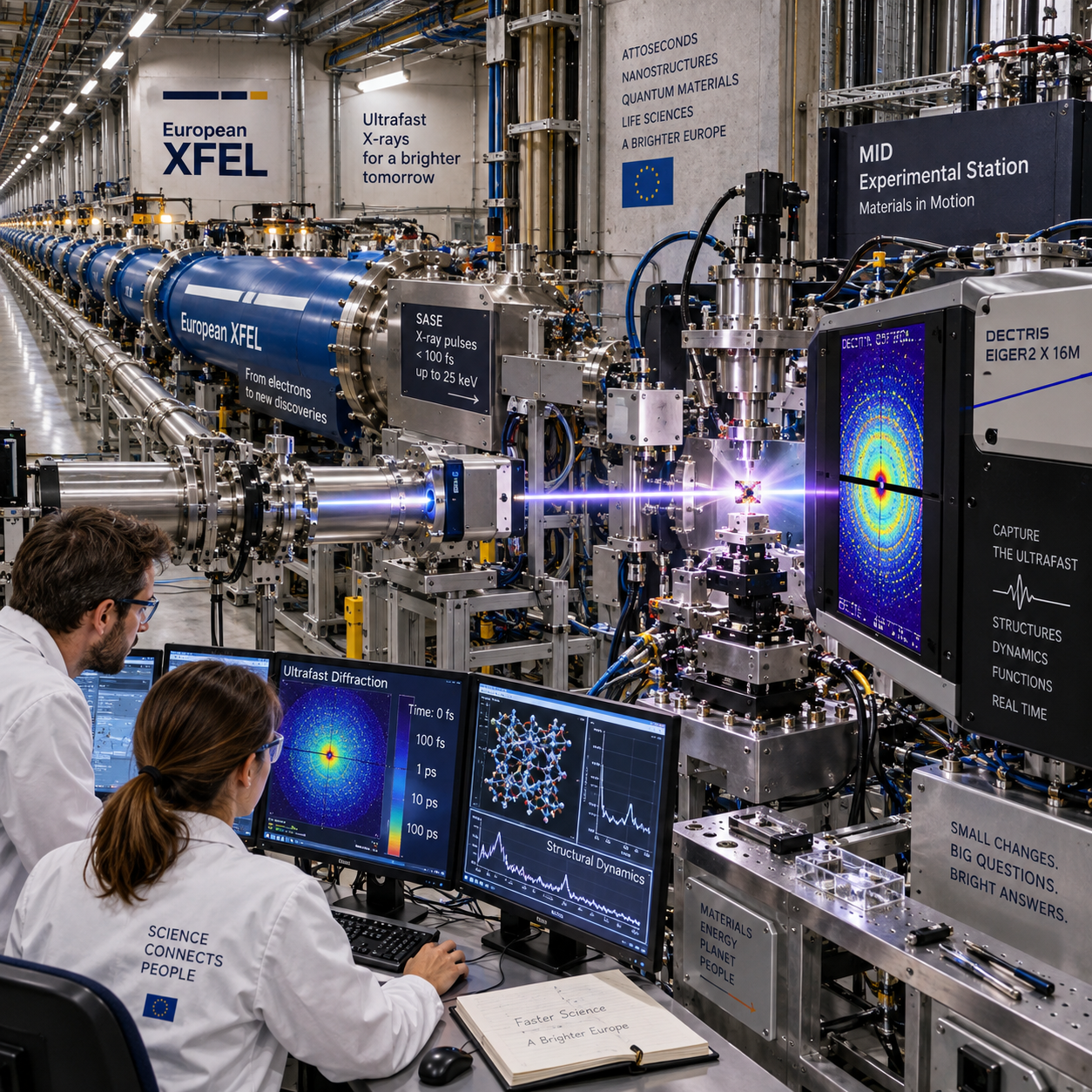
Free-electron lasers (FELs) are changing materials science by allowing researchers to observe structure, chemistry, and function on the timescales when materials actually respond. Their intense, tunable, ultrashort pulses can capture atomic rearrangements, electronic changes, and nanoscale processes that conventional measurements often average together.
That capability matters across energy storage, catalysis, semiconductor technology, magnetism, quantum materials, and advanced manufacturing. FEL experiments do not replace synchrotrons, electron microscopes, laboratory spectroscopy, or simulations. They add a powerful time-resolved view that helps connect a material’s atomic and electronic structure to its real-world behavior.
What Are Free-Electron Lasers?
Free-electron lasers are light sources that generate intense, tunable, ultrashort pulses by passing high-energy electron bunches through an undulator. X-ray free-electron lasers (XFELs) extend this principle into the X-ray range, where the wavelengths are short enough to probe atomic-scale structure.
Unlike a conventional laser, an FEL does not rely on electrons bound inside a fixed material such as a crystal or gas. An accelerator produces a beam of relativistic electrons, and magnetic structures called undulators force the electrons to follow a controlled oscillating path. Their radiation can then be amplified into highly bright pulses.
Researchers can select the photon energy to suit an experiment. This tunability supports diffraction, spectroscopy, imaging, and scattering measurements, while pulse durations reaching the femtosecond range make it possible to investigate ultrafast dynamics.
The practical value comes from combining several beam properties:
- High brightness: many photons arrive in a very short exposure, enabling measurements from small or weakly scattering samples.
- Coherent X-ray pulses: the organized wavefront supports advanced imaging, coherent diffraction, and sensitivity to nanoscale arrangements.
- Short duration: femtosecond pulses can freeze rapid atomic, electronic, and magnetic processes.
- Tunable energy: researchers can target elemental absorption edges and chemical states.
Why FELs Are Transforming Materials Analysis
FELs transform materials analysis by combining high brightness, coherence, elemental sensitivity, and femtosecond-scale time resolution in one experiment. This combination lets researchers measure both what a material looks like and how it changes after excitation.
Many materials-analysis techniques provide excellent structural or chemical information, but they often face a compromise between spatial resolution, temporal resolution, sample size, or sensitivity. A conventional synchrotron can deliver highly valuable measurements with greater routine access and stability. An XFEL adds exceptional peak intensity and time resolution, which is especially useful when a process lasts for picoseconds or femtoseconds.
Element-specific spectroscopy can distinguish how particular atoms participate in a reaction. Diffraction can reveal changes in crystal arrangement. Coherent imaging can expose nanoscale domains, strain, and defects. When these measurements are synchronized with an optical, electrical, or mechanical stimulus, researchers can build a causal sequence rather than simply compare a material before and after treatment.
There are trade-offs. FEL pulses can damage sensitive samples, and the data may require sophisticated models and high-performance computing. The source is therefore most valuable when the research question genuinely depends on transient behavior, weak signals, or difficult sample environments.
Detailed explanations of accelerator-based X-ray sources are available through the U.S. Department of Energy Office of Science, while European researchers can consult facility documentation when planning a proposal.
Watching Materials Change in Real Time
Time-resolved FEL experiments reveal how materials evolve by synchronizing an initiating pump pulse with a delayed probing X-ray pulse. By changing the delay between the two pulses, researchers reconstruct a sequence of structural, chemical, or electronic events.
In a typical pump–probe experiment, the pump may be an optical laser pulse, an electrical signal, a terahertz field, a pressure jump, or a chemical reactant. The FEL pulse acts as the probe. Each measurement records a different delay, and the combined dataset produces a time-dependent picture.
This approach can follow:
- Structural phase transitions, including lattice distortions and melting or crystallization.
- Chemical reactions, such as bond formation, bond breaking, and changes in oxidation state.
- Magnetic switching and the movement of spin or magnetic domains.
- Charge transfer, screening, and other electronic processes in solids.
- Strain propagation and defect formation after mechanical or optical excitation.
The central advance is temporal ordering. A material may appear unchanged in a static measurement even though a short-lived intermediate state controls its final performance. FEL measurements can identify that intermediate state and show whether structure, chemistry, or electronic order changes first.
Good timing is only one part of the experiment. Researchers must also characterize pulse arrival, manage timing jitter, select an appropriate sample environment, and collect enough repeated shots to distinguish a genuine response from fluctuations. A fast measurement with poor synchronization can produce less useful information than a slower, carefully controlled experiment.
Revealing Structure from the Atomic to the Nanoscale
FEL diffraction, spectroscopy, and imaging reveal material structure from atomic arrangements to nanoscale domains, including defects, interfaces, and transient disorder. Each method answers a different part of the structure–function problem.
X-ray diffraction measures how radiation scatters from an ordered lattice. Changes in Bragg peaks can indicate altered lattice spacing, symmetry, strain, or phase composition. When a pump pulse drives a material out of equilibrium, time-dependent diffraction can show how quickly the crystal responds and whether it passes through a short-lived configuration.
Small-angle X-ray scattering and coherent diffraction are useful for larger-scale organization. They can probe nanoparticles, pores, thin films, magnetic domains, and evolving interfaces. Coherent X-ray imaging can also support lensless reconstruction methods, although reconstruction quality depends on signal strength, sample stability, and computational assumptions.
Spectroscopy adds chemical and electronic specificity. By tuning the photon energy near an absorption edge, an experiment can examine a selected element and its local environment. This is particularly useful in complex materials where several atomic species contribute to the overall response.
The result is a layered view:
- Atomic scale: bond lengths, crystal symmetry, lattice motion, and local coordination.
- Nanoscale: domains, interfaces, grain boundaries, defects, and strain fields.
- Functional scale: ion movement, conductivity, switching, catalytic activity, and mechanical response.
Applications Across Materials Science
FELs support materials research wherever performance depends on rapid structural, chemical, magnetic, or electronic changes. Their strongest contribution is often identifying the mechanism behind a useful property.
Energy materials
In batteries, researchers can study how ions move through electrode lattices, how interfaces form, and how strain accumulates during charging. In photovoltaic and thermoelectric materials, ultrafast measurements can follow charge separation, carrier cooling, and lattice responses. These observations help distinguish a promising design from one that merely performs well under a limited set of conditions.
Catalysts and chemical materials
FEL spectroscopy can track oxidation states and local coordination during a catalytic reaction. That information helps identify active sites and transient intermediates that disappear when the reaction stops. Researchers can then compare experimental results with chemical simulations and improve catalyst composition or operating conditions.
Semiconductors and quantum materials
Semiconductor experiments may examine carrier dynamics, charge-density waves, excitons, and structural changes in thin films. Quantum materials introduce additional questions involving correlated electrons, competing phases, and nonequilibrium states. FEL pulses can perturb these systems and reveal how quickly order forms, collapses, or couples to the lattice.
Magnetic and advanced manufacturing materials
Ultrafast X-ray measurements can follow magnetic switching, domain-wall motion, and spin–lattice coupling. In additively manufactured metals and other advanced manufacturing materials, diffraction can map residual stress, phase evolution, and microstructural changes during heating or processing. These results can guide process optimization, though experiments must reproduce relevant temperatures, pressures, cooling rates, and geometries.
The Role of the European FEL Network
The European FEL network connects major facilities, specialized beamlines, user programs, and international teams so researchers can access capabilities that no single laboratory typically provides. Facilities such as the European XFEL, SwissFEL, FERMI, and other European light sources contribute different photon-energy ranges, pulse characteristics, instruments, and sample environments.
Researchers usually begin by defining a measurement that cannot be answered adequately with existing laboratory or synchrotron methods. They then develop a proposal describing the scientific question, sample, required beam properties, timing scheme, safety considerations, and expected data. Peer review determines whether the experiment receives access, often through research infrastructure programs and international collaboration.
Access is only one part of the network’s value. Beamline scientists, instrument teams, sample-environment specialists, detector experts, data scientists, and theory groups can all influence the experiment. A multidisciplinary team may improve the sample geometry before the beamtime, select a more informative detection mode, or connect the result to atomistic simulations.
A practical user journey often includes:
- Define the materials question and identify the required spatial and temporal resolution.
- Test the sample with laboratory methods or conventional synchrotron measurements.
- Prepare a focused proposal with realistic sample quantities and controls.
- Finalize sample delivery, safety documentation, timing plans, and data pipelines.
- Run the beamline experiment, monitor sample damage, and adjust conditions when needed.
- Process, validate, and interpret the data with facility and external collaborators.
Challenges and the Future of FEL-Based Research
FEL research is powerful but demanding: successful experiments require suitable samples, precise timing, careful controls, advanced data analysis, and access to scarce beamtime. Future improvements will make the workflow faster and more reliable, but they will not remove the need for strong experimental design.
Sample damage is a persistent constraint. A high-intensity pulse can alter, vaporize, or permanently transform a sample, especially when the material is thin, hydrated, nanoscale, or chemically reactive. Serial sample delivery, translation between fresh regions, cryogenic approaches, and lower-dose strategies can help, but each introduces engineering and interpretation challenges.
Data volume is another limitation. Modern detectors can record large numbers of single-shot images, spectra, and metadata. Researchers need robust pipelines for calibration, indexing, sorting, uncertainty estimation, and event selection. Machine learning may accelerate classification and anomaly detection, but physical validation remains essential.
Several developments are likely to expand FEL-based materials science:
- Faster detectors with improved dynamic range and lower noise.
- Automated alignment, sample exchange, and adaptive measurement strategies.
- Integrated workflows linking experiments with simulations in near real time.
- Improved high-throughput methods for comparing compositions or processing conditions.
- More realistic operando environments for batteries, catalysts, devices, and manufacturing processes.
Common planning mistakes include requesting an FEL when a conventional synchrotron already answers the question, underestimating sample damage, and treating data analysis as an afterthought. The correction is a staged workflow: establish the baseline with laboratory or synchrotron methods, define the decisive time-dependent measurement, test sample robustness, and agree on analysis methods before beamtime.
Frequently Asked Questions
What is a free-electron laser?
A free-electron laser is an accelerator-based light source that produces intense, tunable radiation from high-energy electrons passing through an undulator. An X-ray free-electron laser generates ultrashort X-ray pulses for structural, chemical, and dynamic measurements.
How do FELs differ from conventional X-ray sources?
FELs provide exceptionally bright and short pulses, often with strong coherence and tunable photon energy. Conventional synchrotron sources generally offer more routine access, stable operation, and excellent average brightness, so the two approaches are complementary.
What materials can researchers study with FELs?
Researchers study energy materials, semiconductors, catalysts, nanomaterials, magnetic materials, quantum materials, thin films, polymers, and advanced manufacturing materials, provided the sample and environment suit the selected experiment.
Why are ultrafast measurements important in materials science?
Ultrafast measurements reveal transient states and causal sequences that static experiments can miss. They show whether atomic motion, charge transfer, chemical change, or magnetic switching drives a material’s response.
How can researchers access European FEL facilities?
Researchers can apply through facility user programs and peer-reviewed proposal calls. A strong application explains the scientific question, sample requirements, beamline technique, timing needs, controls, safety arrangements, and data-analysis plan. Collaboration with beamline scientists often improves feasibility before submission.
FELs are revolutionizing materials science because they connect structure, chemistry, and function across timescales that were previously difficult to observe. Their impact is greatest when the experiment is sharply designed and integrated with conventional characterization, computation, and multidisciplinary expertise. The technology is demanding, but it gives researchers a direct way to watch materials become the materials they are.